
Mucus has a branding problem. Most of us notice it only when we have a cold, a chest infection or a cough that fills the sink with phlegm. So it is easy to think of mucus as waste – something the lungs produce when they are unwell.
In reality, healthy lungs need mucus every minute of the day. A thin film coats the conducting airways, quietly trapping dust, pollen, microbes and other inhaled material before they can settle deeper in the respiratory tract. Modern respiratory research describes mucociliary clearance as one of the lungs’ most important innate defence mechanisms. When it works, we barely know it is happening.
The remarkable part is that mucus is only half the system. The other half is a microscopic moving carpet of cilia. Together they form a self-cleaning conveyor belt that operates from the nose and larger airways down towards the smaller conducting bronchi.
A carefully engineered gel
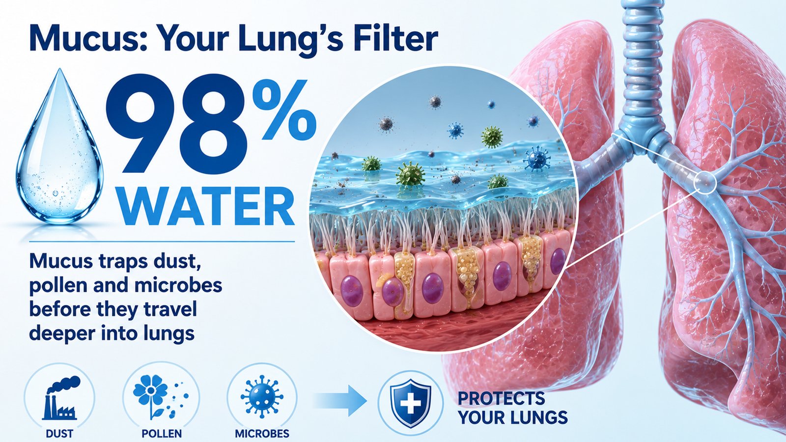
Airway mucus is not simply water mixed with “gunk”. It is a highly organised, viscoelastic gel containing water, salts, proteins, lipids and immune-active molecules. In healthy airways it can be about 98% water, yet the small solid fraction gives it its useful physical properties.
Its key structural ingredients are mucins – enormous, sugar-coated proteins that form a loose molecular mesh. The two main gel-forming mucins in the airways are MUC5B and MUC5AC. In health, MUC5B is usually the more prominent of the two. Mucins are extraordinarily water-loving molecules and can absorb more than 1,000 times their mass in water. That helps explain how a relatively small amount of protein can create a hydrated protective layer.
This gel has to perform a delicate balancing act. It must be sticky enough to capture particles, but fluid enough to move. If it becomes too concentrated, it can become difficult for cilia to propel. If its composition changes, it can become more adhesive, more elastic and more likely to form plugs. The difference between protective mucus and obstructive mucus is therefore often not simply quantity, but quality.
The microscopic escalator in your chest
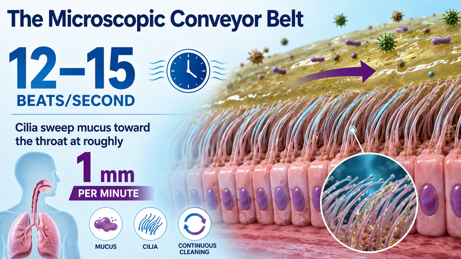
Beneath the mucus lies a thinner watery zone called the periciliary layer. Projecting into it are millions of hair-like cilia attached to airway epithelial cells. These cilia beat in coordinated waves, pushing the mucus towards the throat.
Measurements summarised in a recent physiology review describe cilia beating roughly 12 to 15 times a second, with mucus moving at about 1 mm a minute in healthy airways. The exact speed varies by airway region and conditions, but the principle is the same: particles that entered with one breath are continually being carried back out.
Once secretions reach the upper airway, they are usually swallowed without notice. That may sound unpleasant, but it is normal. The digestive tract then deals with the trapped material. Cough acts as an important back-up system, especially when secretions accumulate in larger airways or the ciliary conveyor belt cannot keep up.
This is why a healthy person does not need to “detox” the lungs with a special syrup. The respiratory tract already possesses a sophisticated cleaning system – provided its mucus stays appropriately hydrated and its cilia remain functional.
Mucus is also part of the immune system
The mucus layer is not merely flypaper. It also carries antimicrobial and immune molecules and creates a physical barrier between inhaled material and the living airway surface. The epithelial cells underneath can recognise danger and release signals that recruit immune cells when infection or injury occurs.
During a viral infection, allergy or exposure to irritants, mucus production may increase. Goblet cells can become more active and inflammatory signals can alter which mucins are produced. This response is meant to protect the airway, but it can overshoot. A defence mechanism that works beautifully in moderation can become a problem when the airways are inflamed and narrowed.
Phlegm colour is often misunderstood too. Yellow or green sputum can occur when large numbers of inflammatory cells enter the mucus. The World Health Organization specifically notes that yellow or green sputum in acute bronchitis does not, by itself, prove a bacterial infection or a need for antibiotics. Colour has to be interpreted alongside fever, breathlessness, duration, examination findings and the patient’s underlying health.
When the conveyor belt breaks
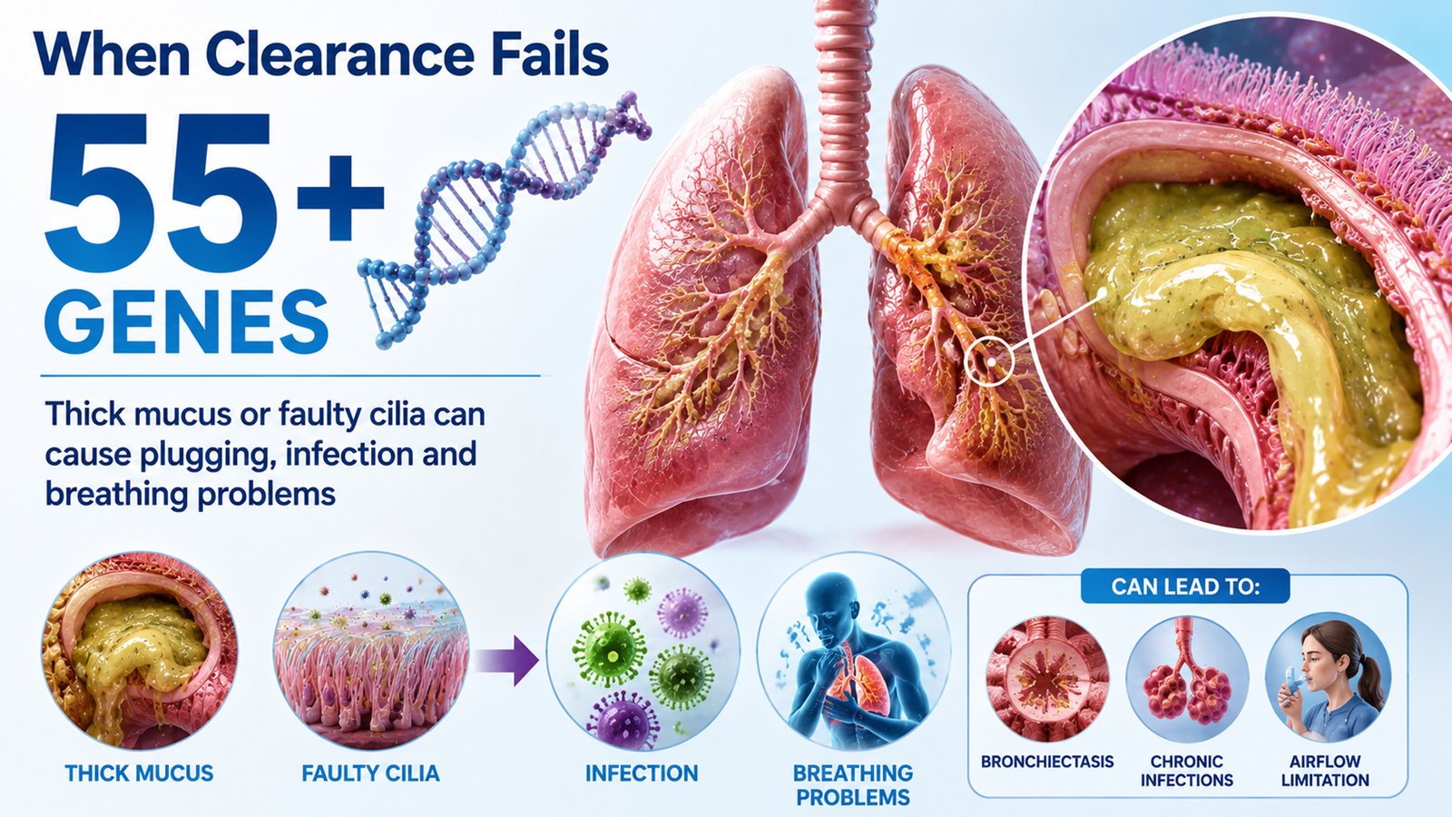
Respiratory disease can disrupt this system at several different points. In asthma, inflammation can drive excessive MUC5AC production and change the balance of airway mucins. Recent experimental work continues to link MUC5AC-rich mucus with impaired mucociliary transport and mucus plugging in asthma. In severe attacks, plugs can block already narrowed airways and contribute to dangerous airflow limitation.
Cystic fibrosis illustrates a different failure. Mutations affecting the CFTR ion channel alter salt and water handling at the airway surface. The result is abnormally concentrated mucus that is difficult to transport, allowing bacteria and inflammatory material to persist. The cilia may be structurally normal, but they are trying to move a gel that has become too dense.
Primary ciliary dyskinesia is almost the mirror image: the mucus may be present, but inherited defects impair the movement of the cilia themselves. The updated joint ERS/ATS diagnostic guideline notes that pathogenic variants in more than 55 genes can cause the condition, which is associated with chronic wet cough, sinus disease and recurrent respiratory problems from poor clearance.
Bronchiectasis can create a vicious cycle of retained secretions, infection and inflammation, which further damages the airways and makes clearance harder. Reflecting how central mucus clearance is to the disease, the 2025 European Respiratory Society guideline strongly recommends teaching airway-clearance techniques to patients with bronchiectasis. These techniques are not generic chest thumping; they are individualised methods, often taught by respiratory physiotherapists, to move secretions more effectively.
Pollution, smoke and the cilia you cannot see
The mucociliary system is also exposed to everything we breathe. That matters in Indian cities where daily life may mean traffic exhaust, construction dust, seasonal air pollution, tobacco smoke or indoor combustion products. The airway lining is the first tissue to meet these exposures.
A 2026 review of airway cilia biology reports that cigarette smoke, PM2.5, allergens and other environmental exposures can impair ciliary structure or function and compromise mucociliary clearance. Smoke can also promote mucus hypersecretion and alter the airway surface environment. In other words, the lungs may face a double burden: more material to clear and a less efficient cleaning mechanism.
Hydration is one of the factors that helps determine whether airway mucus remains transportable. Drinking extra water is not a cure for chronic lung disease, but adequate fluid intake is sensible during febrile illness. Persistent or recurrent sputum deserves attention to the underlying cause rather than simply repeated steam inhalation, cough syrups or antibiotics.
When should phlegm make you seek help?
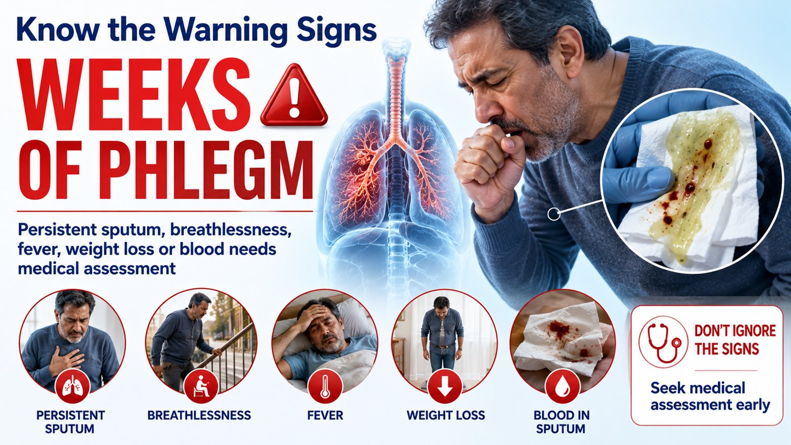
A brief increase in mucus during a cold is common. Medical assessment becomes more important when sputum persists for weeks, repeatedly returns, is associated with breathlessness or wheeze, or is accompanied by fever, weight loss, chest pain or a clear fall in exercise tolerance. In India, a prolonged cough – especially with weight loss, fever, night sweats or blood in the sputum – also requires evaluation for tuberculosis and other significant lung disease.
Coughing up blood should never be dismissed as “just mucus”. Haemoptysis can occur with airway infection, bronchiectasis, tuberculosis, pulmonary embolism or lung cancer, and more than a few streaks – particularly with breathing difficulty or chest pain – needs urgent assessment.
The key is to remember what mucus was designed to do. Its presence is not a failure of the lungs. It is evidence that the lungs possess an active barrier, filtration and transport system. Trouble begins when the gel becomes too thick, too abundant or too adhesive – or when the cilia and cough can no longer move it.
So the next time a doctor asks about phlegm, the question is not simply, “How much mucus is there?” The more useful question is: “Why is it there, and is the airway still able to clear it?” That distinction often tells us far more about what is happening inside the lungs.
This article provides general health information and is not a substitute for individual medical assessment.
(The author is an interventional pulmonologist and respiratory medicine consultant in Bengaluru.)