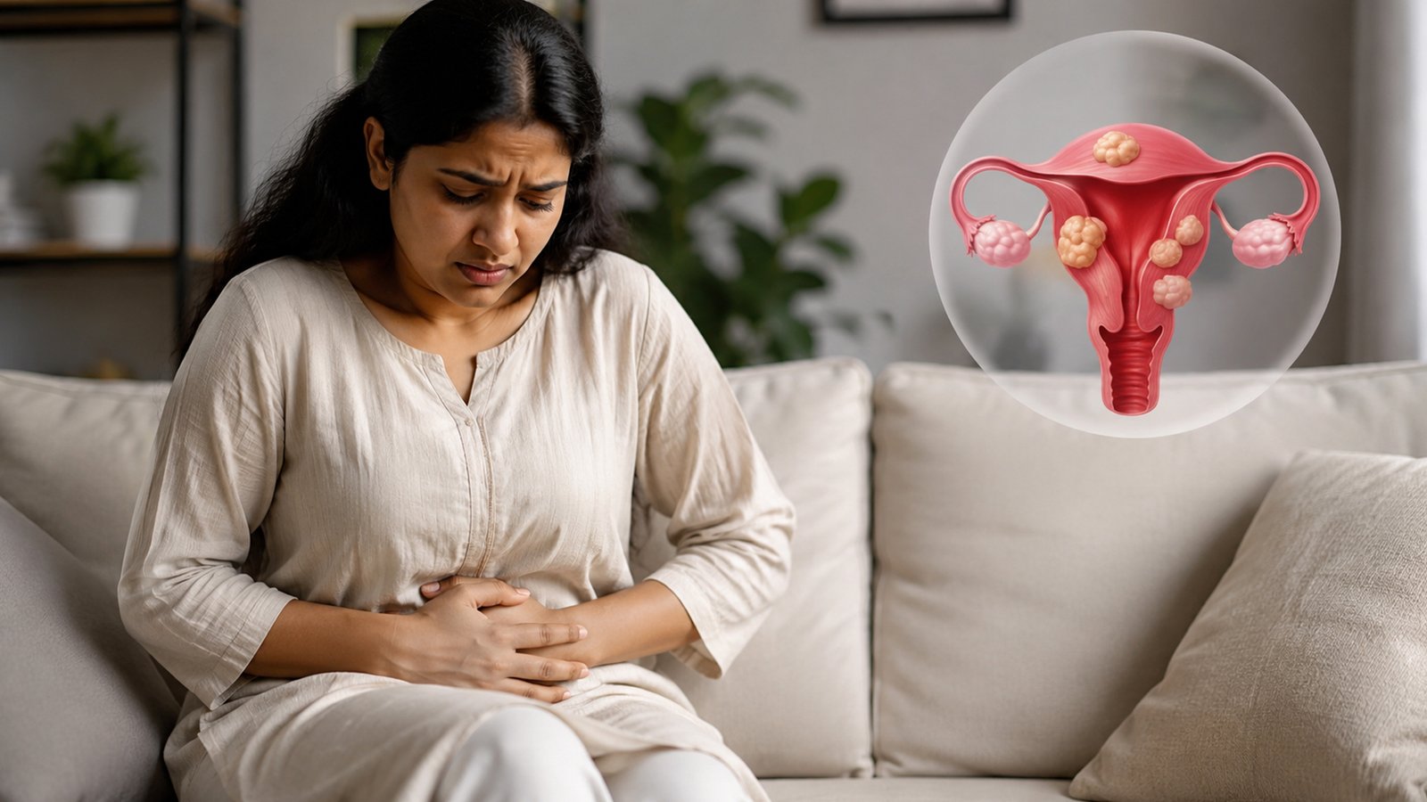
Two women can have the same word on an ultrasound report — “fibroid” — and need completely different advice. One may have a small fibroid on the outer surface of the uterus, feel perfectly well and never need treatment. Another may have a smaller growth that bulges into the uterine cavity and causes flooding periods, iron deficiency and repeated disruption of daily life.
That contrast is the central fact about fibroids. The scan finding itself is not the diagnosis that matters. The real question is whether a fibroid is clinically meaningful: is it plausibly causing the woman’s bleeding, pressure, pain or reproductive problem, and would treating it improve something she actually cares about?
Common does not mean troublesome
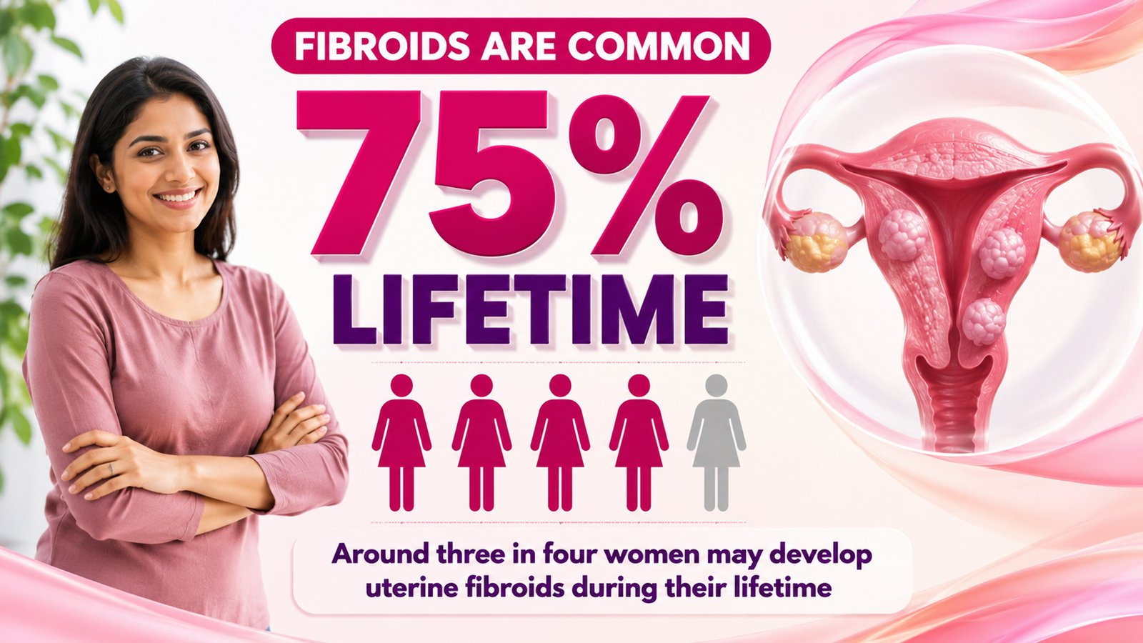
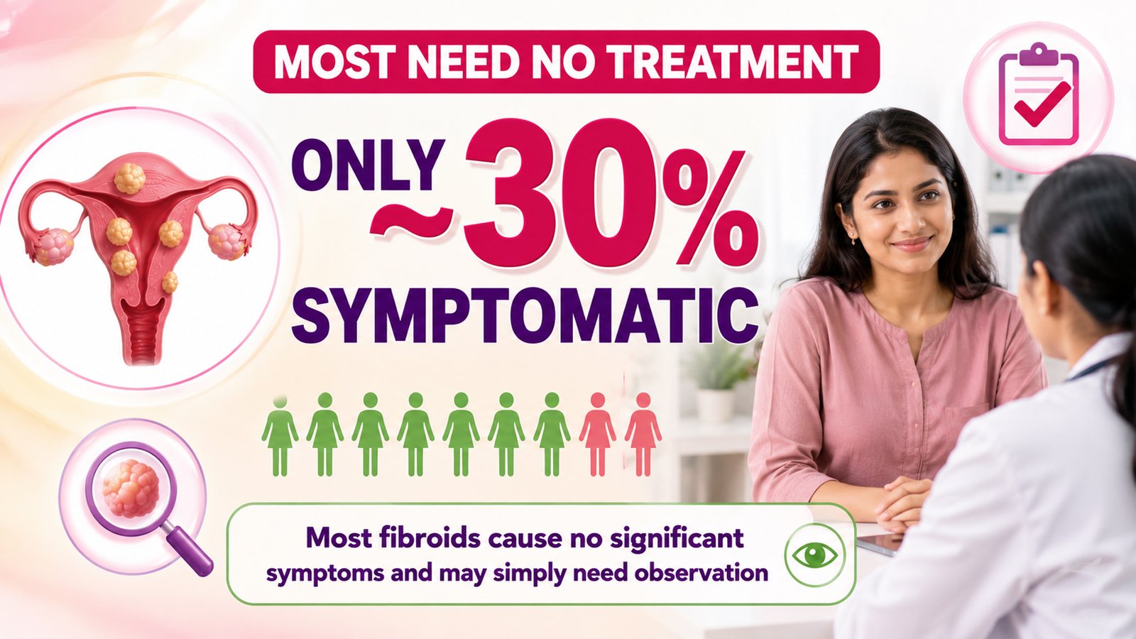
Uterine fibroids, also called leiomyomas or myomas, are benign growths arising from the muscle of the uterus. They are extraordinarily common. Modern FIGO guidance estimates a lifetime prevalence of roughly 75%, yet only about 30% of affected women develop symptoms significant enough to require intervention.
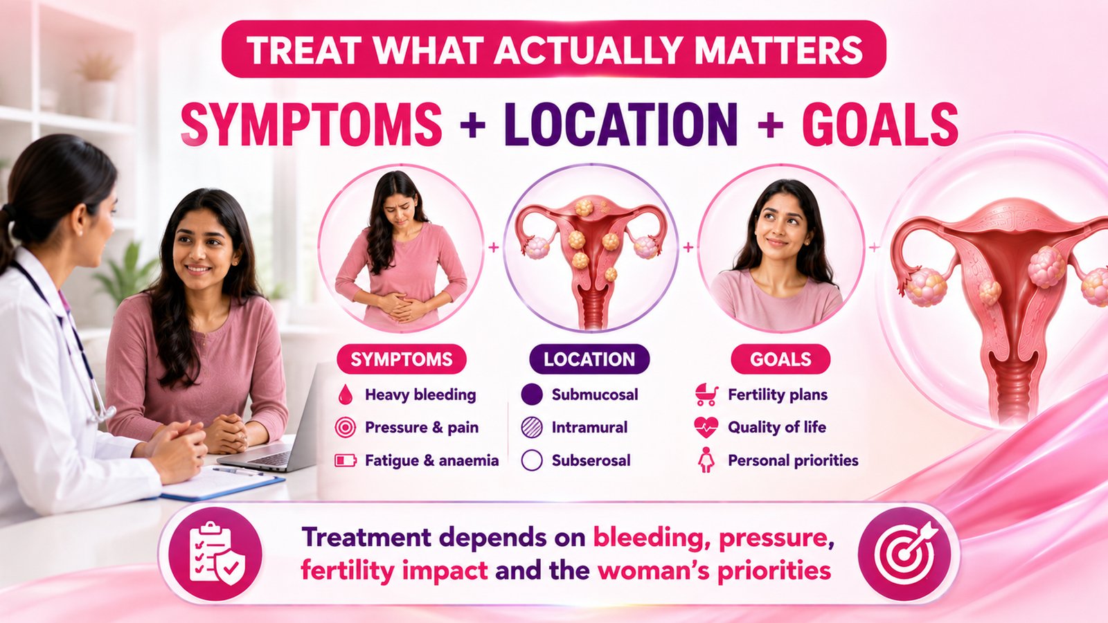
That is why an incidental fibroid is not automatically a problem waiting to happen. Treating every scan finding would expose many women to medicines or procedures without clear benefit. The newest FIGO synthesis, published in August 2026, says management should connect the fibroid’s features with symptoms and the woman’s goals, rather than treating the image in isolation.
THE NUMBER TO REMEMBER | About three in four women may develop fibroids during their lifetime, but most will never need treatment.
Location can matter more than the centimetres
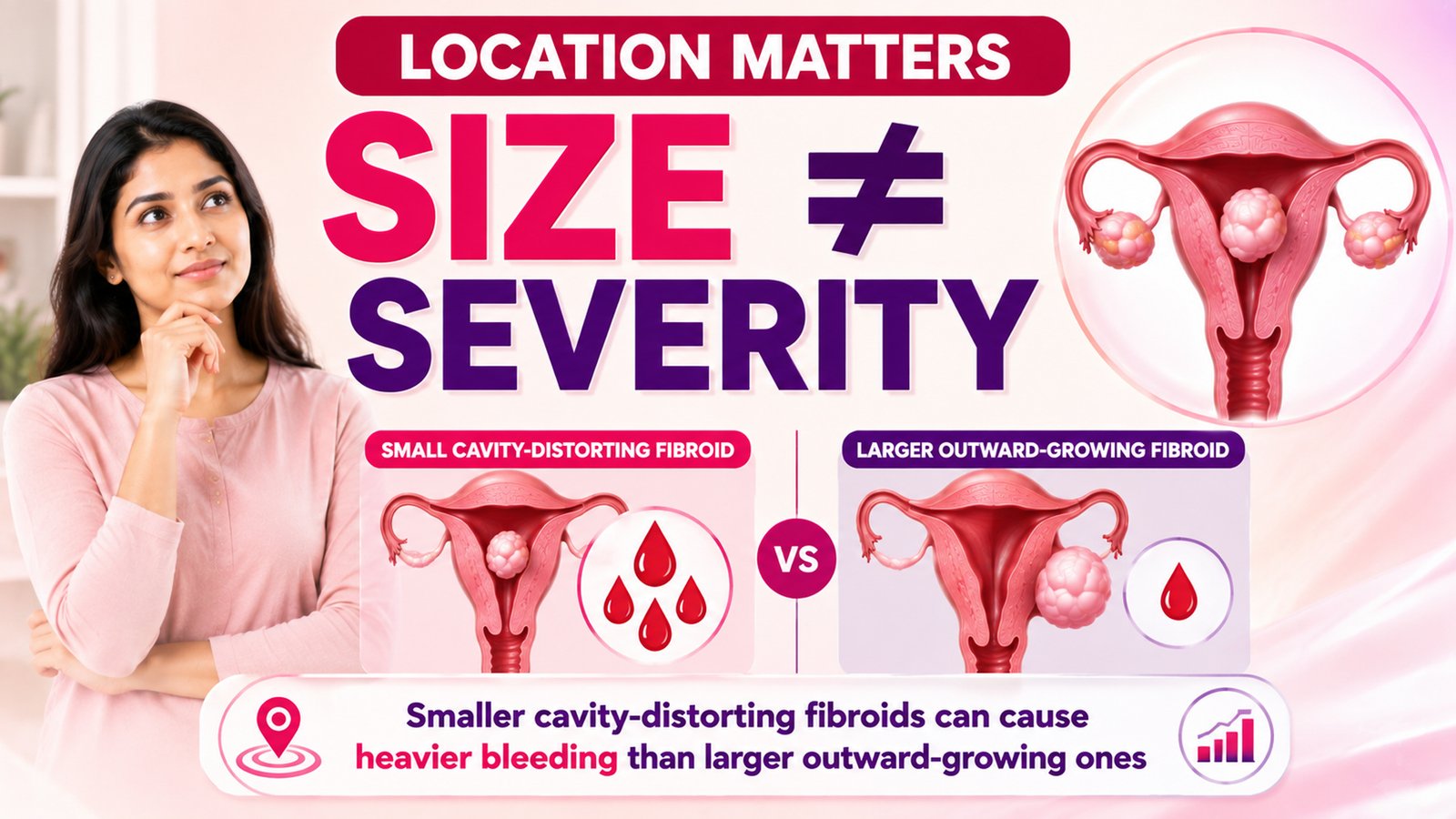
To understand symptoms, it helps to imagine the uterus as a muscular room. A submucosal fibroid pushes towards or into the inner cavity where menstrual bleeding and embryo implantation occur. An intramural fibroid sits within the muscle wall. A subserosal fibroid grows mainly towards the outside of the uterus.
This is why size alone can mislead. A cavity-distorting fibroid may create disproportionately troublesome bleeding or fertility problems, while a larger outward-growing fibroid may produce little bleeding but eventually cause pressure.
Number also matters, as does exactly how close a fibroid lies to the uterine lining, bladder or bowel. Specialists use the FIGO classification to describe this anatomy more precisely than “small”, “medium” or “large”.
Heavy bleeding is the commonest way fibroids announce themselves
Abnormal uterine bleeding is the most frequently reported fibroid symptom. Typical fibroid-related bleeding is often still cyclical — the period arrives when expected — but it is much heavier, lasts longer, or both. Submucosal fibroids have the clearest direct association with heavy menstrual bleeding, probably because they alter the uterine lining, its blood vessels and the local mechanisms that normally stop menstrual bleeding.
The consequence is not merely inconvenient periods. Repeated blood loss can deplete iron and lower haemoglobin, producing iron-deficiency anaemia. Fatigue, palpitations, dizziness and breathlessness may be the symptoms that finally bring a woman to clinic. This matters particularly in India, where the latest WHO country profile estimates that 53.7% of women aged 15–49 were anaemic in 2023. That figure is not an estimate of fibroid-related anaemia; it is a reminder that prolonged heavy bleeding is occurring against an already substantial background burden.
A woman with heavy periods therefore needs more than an ultrasound label. A blood count and, when indicated, iron studies such as ferritin may be useful. Equally, the doctor should not assume that every heavy period is caused by the fibroid: adenomyosis, polyps, thyroid disease, medication effects, pregnancy-related conditions and abnormalities of the uterine lining can coexist.
Pressure symptoms are different from bleeding symptoms
Some fibroids behave less like a bleeding problem and more like a space-occupying one. As the uterus enlarges, women may notice pelvic heaviness, frequent urination and constipation, alongside a lower-abdominal bulge or back discomfort. Anterior fibroids can press on the bladder; posterior ones can press towards the rectum. In severe cases, very large fibroids can obstruct the urinary tract.
These “bulk” symptoms are more closely related to the size, number and position of the growths than to whether they touch the uterine cavity. They are also a good example of why the woman’s experience must lead the decision. A 6 cm fibroid that causes no pressure, bleeding or reproductive issue may reasonably be observed; a similarly sized fibroid that makes a woman urinate every hour or causes constant pelvic fullness may justify treatment.
Fibroids and fertility: avoid blaming the innocent bystander
Finding a fibroid during an infertility work-up is emotionally powerful, but it does not prove causation. The latest FIGO review on fibroids and infertility, published in May 2026, emphasises that location is critical. Submucosal fibroids significantly reduce the chance of pregnancy and can interfere with implantation because they distort or directly influence the cavity. Intramural fibroids may also affect fertility and pregnancy outcomes, but their importance varies with size and proximity to the cavity. Small subserosal fibroids generally have minimal effect on fertility.
This nuance matters. Removing a fibroid is still surgery, with potential bleeding, scarring and recovery. A woman trying to conceive should have the wider fertility picture assessed rather than assuming the fibroid explains everything. When a cavity-involving fibroid is clearly relevant, hysteroscopic myomectomy — performed through the cervix without an abdominal incision — is often the least invasive surgical route for suitable lesions.
How do we decide whether the fibroid is really the culprit?
The first-line imaging test is usually pelvic ultrasound, often combining abdominal and transvaginal views. A good report should describe the number, three-dimensional size and exact location of the important fibroids, and whether they distort the cavity. If the cavity itself needs closer assessment, saline-infusion sonography or hysteroscopy may help. MRI is generally reserved for complex cases or procedural planning, not ordered automatically for every fibroid.
The clinical history is just as important as the scan. When did the bleeding change? Is the cycle still regular? Is there pressure, pain or bladder difficulty? Is pregnancy desired now or later? Has haemoglobin fallen? Depending on age and risk factors, persistent abnormal bleeding may also require sampling of the uterine lining so that a coincidental endometrial problem is not missed.
Treatment should solve a problem, not erase an ultrasound finding
If fibroids are small, asymptomatic or not plausibly responsible for the complaint, observation is often the correct treatment. For women whose main problem is heavy bleeding, medicines can be very effective even though they do not “remove” the fibroid. Options include tranexamic acid, hormonal contraception and progestogen-based treatment. A levonorgestrel-releasing intrauterine system can be useful when the uterine cavity is suitable. Selected GnRH medicines temporarily suppress ovarian hormone signalling and can reduce bleeding and fibroid volume, but choice and duration require medical supervision.
When symptoms are severe, medicines fail, the cavity is mechanically distorted or fertility considerations change the balance, a procedure may be better. Current options include hysteroscopic, laparoscopic or open myomectomy — removal of fibroids while retaining the uterus — and hysterectomy, which removes the uterus and is definitive for women who have completed childbearing and want that option.
Uterine artery embolisation blocks the fibroid’s blood supply, while radiofrequency ablation and high-intensity focused ultrasound are uterus-sparing alternatives for selected women.
Future pregnancy plans must be discussed before choosing among these procedures. The evidence for fertility after newer image-guided techniques is still evolving, and FIGO does not recommend uterine artery embolisation for women actively wishing to conceive. There is no universally “best” fibroid procedure; there is a best-fit option for a particular fibroid pattern, symptom burden and reproductive plan.

When should a fibroid prompt medical review?
Seek assessment for periods that have become persistently very heavy or prolonged, symptoms of anaemia, pelvic pressure that is worsening, difficulty passing urine, unexplained abdominal enlargement, infertility or recurrent pregnancy loss, or pain that is new and significant. Bleeding that is causing faintness, marked breathlessness, chest discomfort or weakness deserves urgent care rather than waiting for a routine appointment.
One cancer fear also deserves perspective. Fibroids are benign tumours, and malignancy in a presumed fibroid is rare. Imaging cannot provide absolute certainty in every unusual mass, however, and the threshold for careful evaluation is higher after menopause or when imaging looks atypical.
The most important measurement is not always on the scan
A fibroid becomes a problem when its presence and position fit the woman’s symptoms, when it causes measurable harm such as anaemia, when it creates meaningful pressure, or when it is likely to interfere with fertility or pregnancy goals. The centimetres help us plan. They do not, by themselves, tell us whether to treat.
For many women, the safest decision is simply to know the fibroid is there and leave it alone. For others, controlling bleeding restores energy; removing a cavity-distorting fibroid may improve reproductive prospects; treating a bulky uterus may give back comfort and bladder space. The job of good fibroid care is not to make every scan look normal.
It is to identify which fibroids actually matter — and treat those, for a reason.
This article provides general health information and is not a substitute for individual medical assessment.
(The author is a consultant obstetrician and gynaecologist based in Bengaluru.)